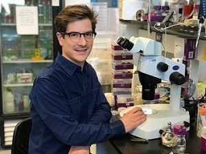
Staff profile

| Affiliation |
|---|
| Assistant Professor in the Department of Biosciences |
Biography
Cell division varies with cell identity
Cytokinesis is the process by which one cell physically divides into two at the end of mitosis. This process is fundamental for life, and errors can result in abnormal chromosomal numbers, developmental defects, and cancers. Work over the last century has shown that cytokinesis in animal cells requires a carefully synchronised set of molecular signals that promote the formation of a contractile ring around the cell equator, which then constricts to divide the cell. Similarities in the structural and molecular organization of this division apparatus in a variety of animal model systems give the impression that the mechanisms underlying cell division do not vary between cell and organism types. However, clinical studies have shown tissue-specific division failure due to mutations in cytokinetic proteins. In addition to this, we have shown that in the early Caenorhabditis elegans embryo the requirement for an ‘essential’ cytokinesis protein varies between specific cells. These results highlight an under-appreciated intersection between cell identity and cell division. Therefore, our lab is expanding work in this area, analysing cell division in a multicellular context to identify how cells from the same organism are regulated in different ways during cytokinesis.
C. elegans is an ideal tool to study cell division
Caenorhabditis elegans is a small (1mm) nematode worm that has several characteristics that make it an excellent model in which to investigate context-specific variation in cell division. First, it has a stereotyped development in which each cell has a different identity, allowing cytokinetic phenotypes to be compared between different cells. Second, C. elegans has been used as a model system for decades and many of the pathways that contribute to specifying cell fate during development are well known. Third, genetic tools allow us to precisely disrupt or modify specific protein function. Fourth, cell division and embryogenesis can be observed using live-cell fluorescence microscopy, allowing analysis and quantification of these dynamic processes. Finally, many pathways involved in cytokinesis and cell identity are highly conserved between C. elegans and higher metazoans, which enables comparison with other model systems. Taking advantage of these features, our lab currently investigates how cytokinesis varies between cells in the early C. elegans embryo using a combination of genetics and fluorescence microscopy.
Research interests
- C. elegans development
- Cell division
- Cytoskeletal structure and dynamics
- Live-cell fluorescence microscopy
Publications
Chapter in book
- Using fast-acting temperature-sensitive mutants to study cell division in Caenorhabditis elegansDavies, T., Sundaramoorthy, S., Jordan, S., Shirasu-Hiza, M., Dumont, J., & Canman, J. (2017). Using fast-acting temperature-sensitive mutants to study cell division in Caenorhabditis elegans. In A. Echard (Ed.), Methods in Cell Biology. (pp. 283-306). Elsevier. https://doi.org/10.1016/bs.mcb.2016.05.004
Edited book
- Landslide Hazards, Risks, and DisastersDavies, T., Rosser, N., & Shroder, J. (Eds.). (2022). Landslide Hazards, Risks, and Disasters. Elsevier. https://doi.org/10.1016/c2018-0-02502-5
Journal Article
- Mechanical coordination between anaphase A and B drives asymmetric chromosome segregationDias Maia Henriques, A. M., Davies, T. R., Dmitrieff, S., Minc, N., Canman, J. C., Dumont, J., & Maton, G. (2026). Mechanical coordination between anaphase A and B drives asymmetric chromosome segregation. Journal of Cell Biology, 225(1), Article e202505038. https://doi.org/10.1083/jcb.202505038
- Germ fate determinants protect germ precursor cell division by reducing septin and anillin levels at the cell division plane.Connors, C. Q., Mauro, M. S., Wiles, J. T., Countryman, A. D., Martin, S. L., Lacroix, B., Shirasu-Hiza, M., Dumont, J., Kasza, K. E., Davies, T. R., & Canman, J. C. (2024). Germ fate determinants protect germ precursor cell division by reducing septin and anillin levels at the cell division plane. Molecular Biology of the Cell, 35(7), Article mbcE24020096T. https://doi.org/10.1091/mbc.E24-02-0096-T
- Oligodendroglial macroautophagy is essential for myelin sheath turnover to prevent neurodegeneration and deathAber, E. R., Griffey, C. J., Davies, T., Li, A. M., Yang, Y. J., Croce, K. R., Goldman, J. E., Grutzendler, J., Canman, J. C., & Yamamoto, A. (2022). Oligodendroglial macroautophagy is essential for myelin sheath turnover to prevent neurodegeneration and death. Cell Reports, 41(3), Article 111480. https://doi.org/10.1016/j.celrep.2022.111480
- CYK4 relaxes the bias in the off-axis motion by MKLP1 kinesin-6Maruyama, Y., Sugawa, M., Yamaguchi, S., Davies, T., Osaki, T., Kobayashi, T., Yamagishi, M., Takeuchi, S., Mishima, M., & Yajima, J. (2021). CYK4 relaxes the bias in the off-axis motion by MKLP1 kinesin-6. Communications Biology, 4, Article 180. https://doi.org/10.1038/s42003-021-01704-2
- FHOD‐1 is the only formin in Caenorhabditis elegans that promotes striated muscle growth and Z‐line organization in a cell autonomous mannerSundaramurthy, S., Votra, S., Laszlo, A., Davies, T., & Pruyne, D. (2020). FHOD‐1 is the only formin in Caenorhabditis elegans that promotes striated muscle growth and Z‐line organization in a cell autonomous manner. Cytoskeleton, 77(10), 422-441. https://doi.org/10.1002/cm.21639
- Cell-intrinsic and -extrinsic mechanisms promote cell-type-specific cytokinetic diversityDavies, T., Kim, H. X., Romano Spica, N., Lesea-Pringle, B. J., Dumont, J., Shirasu-Hiza, M., & Canman, J. C. (2018). Cell-intrinsic and -extrinsic mechanisms promote cell-type-specific cytokinetic diversity. ELife, 7, Article e36204. https://doi.org/10.7554/elife.36204
- FLIRT: fast local infrared thermogenetics for subcellular control of protein functionHirsch, S. M., Sundaramoorthy, S., Davies, T., Zhuravlev, Y., Waters, J. C., Shirasu-Hiza, M., Dumont, J., & Canman, J. C. (2018). FLIRT: fast local infrared thermogenetics for subcellular control of protein function. Nature Methods, 15(11), 921-923. https://doi.org/10.1038/s41592-018-0168-y
- Low Efficiency Upconversion Nanoparticles for High-Resolution Coalignment of Near-Infrared and Visible Light Paths on a Light MicroscopeSundaramoorthy, S., Garcia Badaracco, A., Hirsch, S. M., Park, J. H., Davies, T., Dumont, J., Shirasu-Hiza, M., Kummel, A. C., & Canman, J. C. (2017). Low Efficiency Upconversion Nanoparticles for High-Resolution Coalignment of Near-Infrared and Visible Light Paths on a Light Microscope. ACS Applied Materials and Interfaces, 9(9), 7929-7940. https://doi.org/10.1021/acsami.6b15322
- Cell polarity is on PAR with cytokinesisDavies, T., Jordan, S. N., & Canman, J. C. (2016). Cell polarity is on PAR with cytokinesis. Cell Cycle, 15(10), 1307-1308. https://doi.org/10.1080/15384101.2016.1160667
- Cortical PAR polarity proteins promote robust cytokinesis during asymmetric cell divisionJordan, S. N., Davies, T., Zhuravlev, Y., Dumont, J., Shirasu-Hiza, M., & Canman, J. C. (2016). Cortical PAR polarity proteins promote robust cytokinesis during asymmetric cell division. Journal of Cell Biology, 212(1), 39-49. https://doi.org/10.1083/jcb.201510063
- CYK4 Promotes Antiparallel Microtubule Bundling by Optimizing MKLP1 Neck ConformationDavies, T., Kodera, N., Kaminski Schierle, G. S., Rees, E., Erdelyi, M., Kaminski, C. F., Ando, T., & Mishima, M. (2015). CYK4 Promotes Antiparallel Microtubule Bundling by Optimizing MKLP1 Neck Conformation. PLoS Biology, 13(4), Article e1002121. https://doi.org/10.1371/journal.pbio.1002121
- High-Resolution Temporal Analysis Reveals a Functional Timeline for the Molecular Regulation of CytokinesisDavies, T., Jordan, S., Chand, V., Sees, J., Laband, K., Carvalho, A. X., Shirasu-Hiza, M., Kovar, D., Dumont, J., & Canman, J. (2014). High-Resolution Temporal Analysis Reveals a Functional Timeline for the Molecular Regulation of Cytokinesis. Developmental Cell, 30(2). https://doi.org/10.1016/j.devcel.2014.05.009
- Stuck in the middle: Rac, adhesion, and cytokinesisDavies, T., & Canman, J. C. (2012). Stuck in the middle: Rac, adhesion, and cytokinesis. Journal of Cell Biology, 198(5). https://doi.org/10.1083/jcb.201207197
- Cytokinesis microtubule organisers at a glanceLee, K., Davies, T., & Mishima, M. (2012). Cytokinesis microtubule organisers at a glance. Journal of Cell Science, 125(15). https://doi.org/10.1242/jcs.094672
- Aurora B and 14-3-3 Coordinately Regulate Clustering of Centralspindlin during CytokinesisDouglas, M. E., Davies, T., Joseph, N., & Mishima, M. (2010). Aurora B and 14-3-3 Coordinately Regulate Clustering of Centralspindlin during Cytokinesis. Current Biology, 20(10). https://doi.org/10.1016/j.cub.2010.03.055
- Drosophila PAT1 is required for Kinesin-1 to transport cargo and to maximize its motilityLoiseau, P., Davies, T., Williams, L., Mishima, M., & Palacios, I. (2010). Drosophila PAT1 is required for Kinesin-1 to transport cargo and to maximize its motility. Development., 137(16). https://doi.org/10.1242/dev.048108

