Alan Reece X-Ray Imaging Laboratory

Welcome to the XCT Laboratory of Durham University Engineering

The X-ray Computed Tomography (XCT) Laboratory at Durham Engineering gives researchers and industry partners access to cutting-edge 3D imaging technology. From microstructural analysis of advanced materials to non-destructive inspection of industrial components, our facility delivers high-resolution internal imaging. Whether you're new to XCT or an experienced user looking for the right scanner, you'll find everything you need here.
Learn more about us
Measurement Lab
Scanned rock sample

Discover What XCT Can Do For You

X-ray Computed Tomography reveals the hidden interior of your samples in stunning 3D detail, completely non-destructive. Whether you're characterising microstructures or inspecting industrial components, XCT delivers answers without damaging what matters.

Learn more about X-Rays
RX cabin

Our Machines

We host two complementary scanners to match any application. The EasyTom L handles larger samples with faster acquisition, while the Xradia Versa 410 delivers sub-micron resolution for detailed microstructural work. Together, they cover the full range of XCT imaging needs.

Discover Our Machines

Gallery of Example Scans


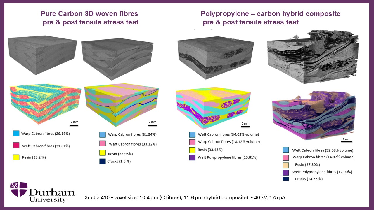
X-ray CT enables non-destructive characterisation of composite materials by revealing their internal architecture in three dimensions, including fibre orientation, resin distribution, porosity, and damage development. By comparing specimens before and after mechanical testing, XCT can quantify crack formation, delamination, fibre rearrangement, and other failure mechanisms that govern material performance. These capabilities support materials development, quality assurance, and structural integrity assessments across applications ranging from aerospace and energy systems to automotive and civil engineering composites.


Scan with us
Composite material under XCT


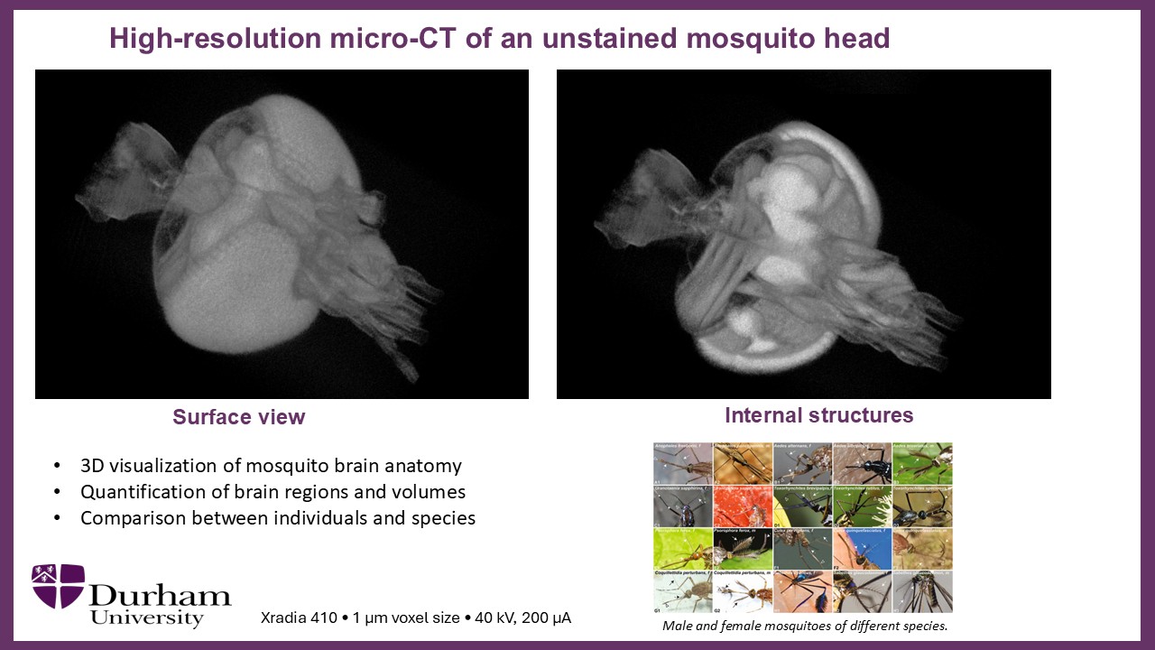
Mosquito head scanned at 1 µm voxel resolution using high-resolution micro-CT, revealing both external morphology and internal anatomical structures without dissection or staining. The scan enables detailed 3D visualisation and quantitative analysis of features such as sensory organs and brain regions. These data support comparative studies across individuals and species while preserving the specimen intact.


Scan with us
Mosquito head under XCT


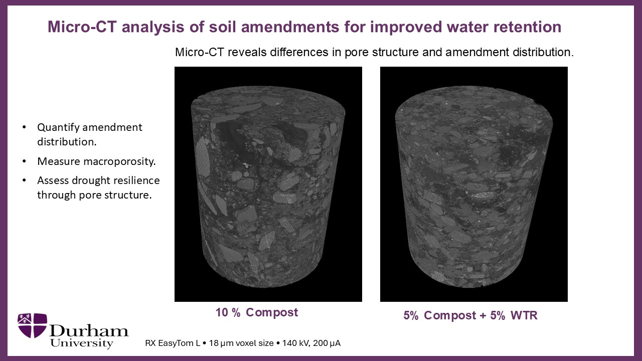
Soil cores scanned at 18 µm voxel resolution using micro-CT to visualise the three-dimensional distribution of soil amendments and pore networks. The scans enable quantitative measurement of porosity, amendment mixing, and structural heterogeneity that influence water retention and transport. These insights support the development and optimisation of soils with improved drought resilience and hydraulic performance.


Scan with us
Soil sample
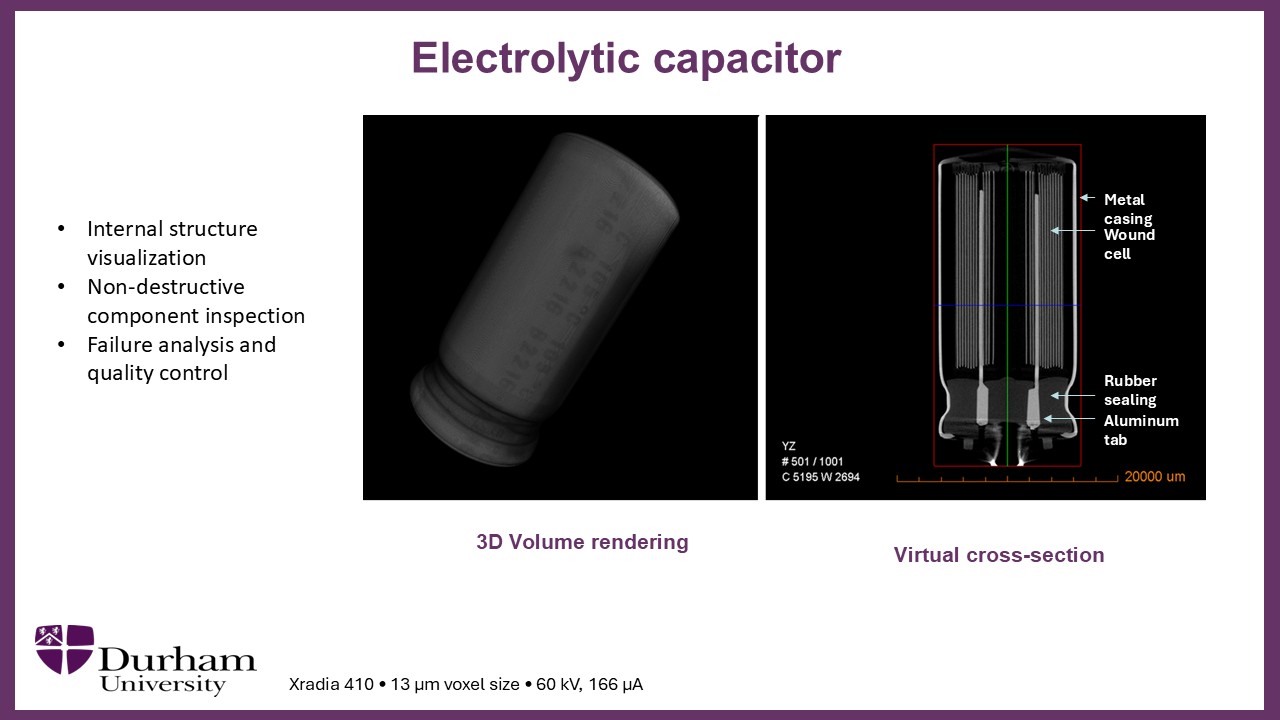
X-ray CT provides a powerful non-destructive method for inspecting the internal structure of electronic components, from individual capacitors and connectors to complete assemblies and consumer devices. Differences in material density create striking contrast in X-ray images, allowing internal features such as conductors, solder joints, insulation layers, casings, and mechanical components to be visualised in exceptional detail. These datasets support quality control, defect detection, failure analysis, reverse engineering, and reliability studies without disassembly, preserving valuable or operational equipment intact.


Scan with us
Capacitor sample under XCT
Two students walking past a coffee shop in Durham City centre

Access and How to Apply

The Durham Engineering XCT Laboratory is open to academic collaborators, industry partners, and postgraduate students. Scan time is available through multiple routes, discover the best option for you!.

How to apply for scan time
Three students walking with a flight of stairs in the background

Meet the team

Our lab is run by a dedicated team of engineers and researchers with expertise spanning XCT and a wide range of applications, from geotechnics to biomedical science. Whatever your sample or question, we're here to help.

Meet the Team